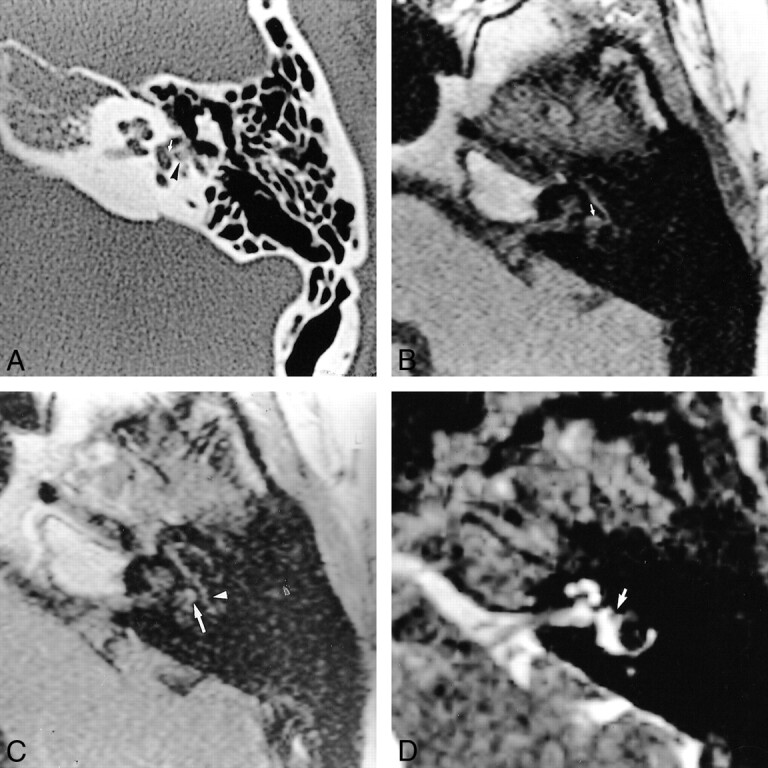
fig 4.

44-year-old woman referred for SNHL and vertigo 6 weeks after stapedectomy with a granuloma located around the prosthesis and in the oval window niche.
A, Axial CT scan of the left temporal bone shows well-located prosthesis (arrow) and partial filling of the posterior part of the oval window niche (arrowhead).
B, Noncontrast axial T1-weighted MR image (500/15) shows normal signal intensity of the labyrinthine fluid, except for slightly high intensity around the oval window (arrow).
C, Contrast-enhanced axial T1-weighted MR image (500/15) shows mild enhancement of the oval window (arrow). Tympanic portion of the facial nerve is seen laterally (arrowhead).
D, Axial T2-weighted MR image shows normal high signal intensity of the labyrinthine fluid. A discrete intravestibular bulging of the prosthesis is clearly depicted (arrow).
