fig 2.
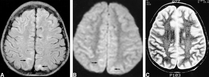
Images of a 12-year-old male patient (patient 2) with generalized tonicoclonic seizure.
A, Initial FLAIR image shows increased signal intensity in the cortical gray matter and subcortical white matter in cuneus and precuneus bilaterally (arrows).
B, Initial diffusion-weighted image shows mildly increased signal intensity in the corresponding areas (arrows). The decrease of the mean ADC was 8% at the right and 6% at the left on the ADC map (not shown).
C, Follow-up T2-weighted image obtained 14 days after the onset of seizure shows complete resolution of the signal change.

