
fig 3.
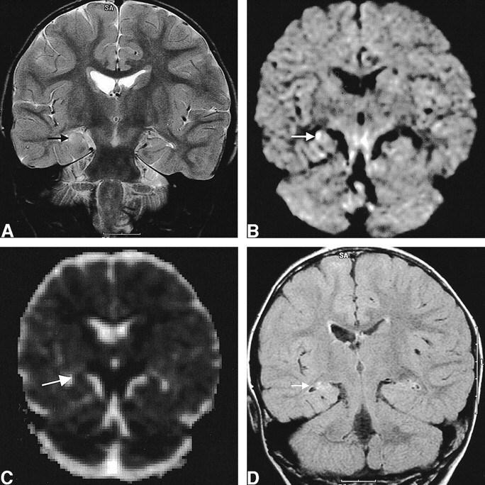
Images of a 2-year-old female patient (patient 6) with complex partial status epilepticus with secondary generalization.
A, Initial T2-weighted image shows increased signal intensity and swelling in the right hippocampus (arrow).
B, Initial diffusion-weighted image shows increased signal intensity in the right hippocampus (arrow).
C, Corresponding ADC map shows 14% decrease of mean ADC (arrow).
D, Follow-up FLAIR image obtained 18 months after the onset of seizure shows the resolution of the swelling and mass effect of the hippocampus (arrow) and increased T2 signal intensity without definite atrophic change of the hippocampus.