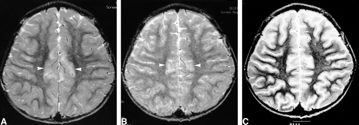
fig 6.
Images of a 3-year-old male patient (patient 1) with complex partial status epilepticus show the resolution process of the signal change.
A, Initial T2-weighted image shows increased signal intensity in the cortical gray matter of bilateral cingulate gyri (arrows).
B, Follow-up T2-weighted image shows partial resolution of the signal intensity 9 days after seizure onset.
C, Follow-up T2-weighted image shows complete resolution of the signal intensity 30 days after seizure onset.

