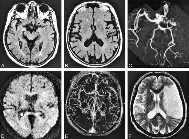
fig 1.

A 78-year-old woman who underwent imaging 35 minutes after onset of right hemiparesis and aphasia.
A and B, FLAIR images (8000/10/1 [TR/TE/excitation]), TI = 2000) show contiguous intraarterial signal, which is hyperintense to cerebral parenchyma in M1, M2, and M3 segments of the left middle cerebral artery (arrows).
C, MR angiogram (32/6.8, flip angle = 15 degrees) shows corresponding lack of TOF effect in M1, M2, and M3 segments of the left middle cerebral artery.
D, Diffusion-weighted image (500/123/1, b = 1200) shows faintly hyperintense lesion localized in the area fed by left lateral lenticulostriate artery (arrow); however, there is no change in the hemispheric territory of the left middle cerebral artery.
E, Postcontrast MR angiogram (32/6.8, flip angle = 15 degrees) shows intraluminal enhancement in the left middle cerebral artery except for in the distal potion of the M1 segment. Arrow indicates complete obstruction in distal portion of left M1 segment.
F, T2-weighted image (4500/96/1) obtained 7 days after onset confirmed final infarction is in the entire territory of the left middle cerebral artery. Note the diffusion-weighted lesion is still smaller than the area of intraarterial signal distribution. Intraarterial signal consists of not only complete obstruction but also slow flow. The area of final infarct corresponds to the area of intraarterial signal distribution.
