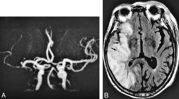
fig 4.
An 83-year-old woman who underwent imaging 23.5 hours after onset of loss of consciousness.
A, MR angiogram (32/6.8, flip angle = 15 degree) shows occlusion in distal portion of M1 segment of the right middle cerebral artery.
B, FLAIR image (8000/10/1, TI = 2000) already shows hyperintense lesion and cortical swelling in the right middle cerebral artery territory. Intraarterial signal (arrows) is shown in the right middle cerebral artery corresponding to lack of TOF; however, signal is hampered by narrowed sulci owing to vasogenic edema.

