fig 5.
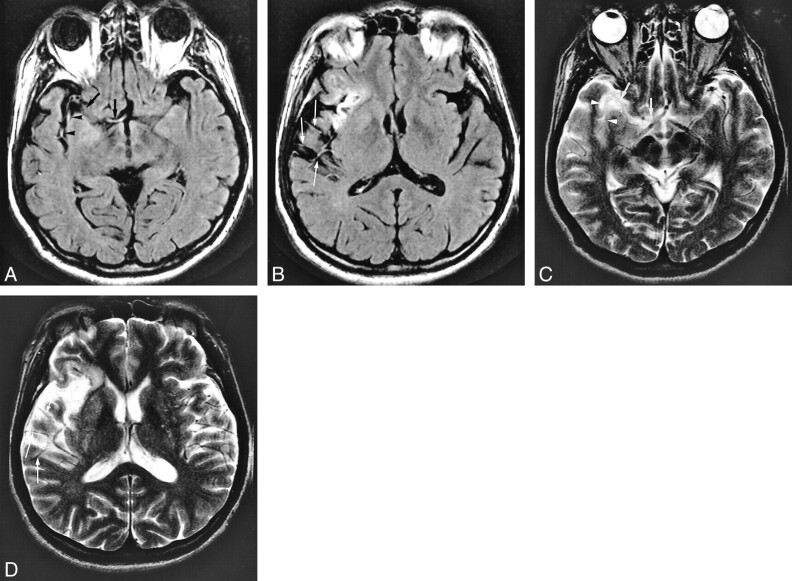
A 62-year-old man who underwent imaging 2.5 hours after onset of loss of consciousness and left hemiparesis.
A and B, FLAIR images (8000/10/1, TI = 2000) show intraarterial signal in the M1 (arrows), M2 (arrowheads), and M3 (thin long arrows) segments of the right middle cerebral artery. Old infarction proceeds in the right insular cortex.
C and D (same level as in A and B), T2-weighted images (4500/96/1) show lack of flow void in M1 (arrows) and M2 (arrowheads) segments; however, evidence of flow void is visible in the M3 segment (thin long arrows).
MR angiogram (not shown) shows lack of TOF in the right middle cerebral artery. CT confirmed final infarction in both perforator and hemispheric-branch territories of the right middle cerebral artery.

