fig 3.

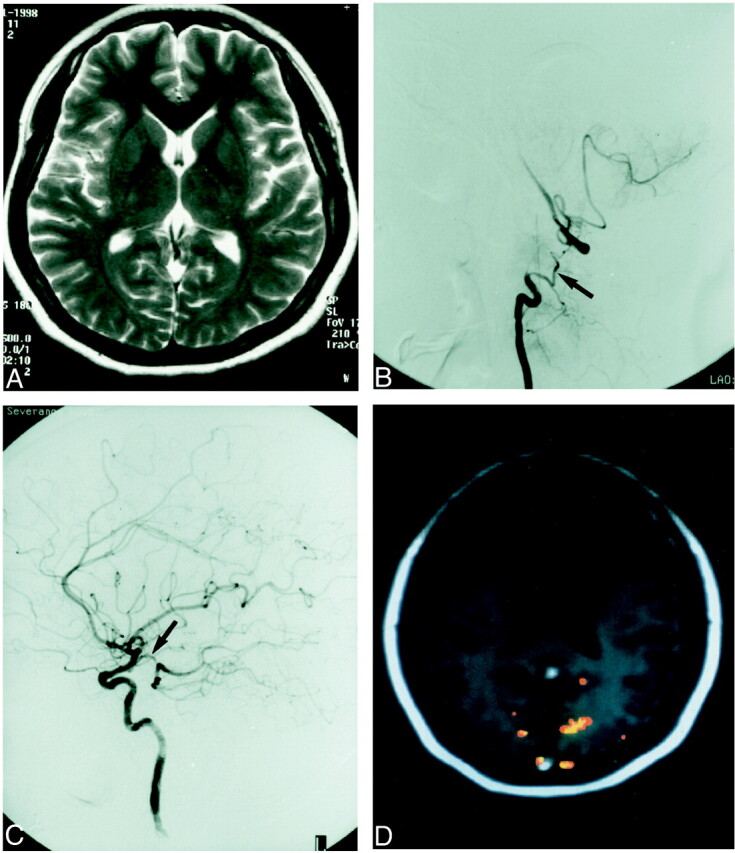
A 29-year-old woman (patient 3) with intermittent left homonymous hemianopsia.
A, T2-weighted image [4500/120 (TR/TE)] is normal.
B and C, Digital subtraction angiogram (B) shows discontinuity of the right vertebral artery and collateral vessels (arrow). The clinical and angiographic diagnosis was dissection of the vertebrobasilar artery. The right posterior communicating artery was hypoplastic (C), whereas the left posterior communicating artery (arrow) showed good patency. The clinical symptom of visual problem in this patient was believed to appear when blood flow from the basilar artery decreased.
D, fMR [90/56/40° (TR/TE/flip angle)] shows decreased activity in the right visual cortex.
