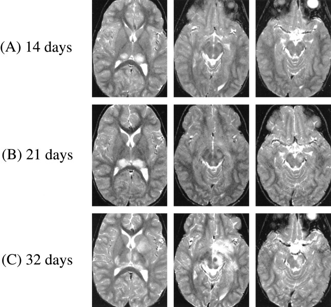
fig 1.
A–C. T2-weighted MR images at 3000/100/1 (TR/TE/excitation) at the following times after initial symptom (headache) onset. A, At 14 days, bilateral thalamic lesions (left slightly more prominent than right). B, At 21 days, thalamic lesions have increased in size and new lesion has appeared in left cerebral peduncle (lesions in left globus pallidus and forceps major not shown). C, At 32 days, further progression of thalamic lesions to involve right geniculate body, right posterior limb of internal capsule, and complete involvement of left putamen and globus pallidus tracking down to left cerebral peduncle (bilateral lesions in hemispheric white matter and left frontoparietal cortex not shown)

