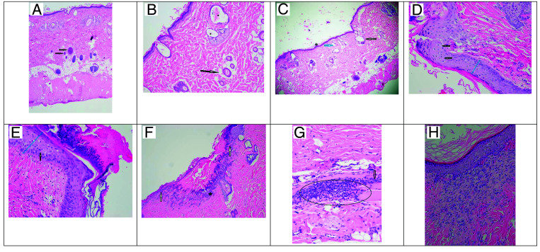
Figure 1.
Dermal pathology on Days 0, 3, and 10. (A) Normal full thickness intact skin with the black arrows indicating normal hair follicles (40× magnification). (B) Day 0 Depilatory method with the arrow indicating a normal hair follicle with central hair shaft. The asterisks (*) indicate hair follicles that are moderately dilated with hair shaft fragments, flattened follicular epithelium, and compressed sebaceous glands (10× magnification). (C) Day 0 Clipper method with the arrow indicating a markedly dilated hair follicle void of the hair shaft and a compressed sebaceous gland. The blue arrow indicates a moderately dilated hair follicle with no hair shaft (40× magnification). (D) Day 3 Clipper method shows mildly transmural thickened epidermis (blue line) and the arrow shows single cell necrosis (20× magnification). (E) Day 3 Depilatory method shows a moderate transmural thickened epidermis (blue line) with the arrow showing cellular bridging (edema). A thick mat of serocellular debris is seen covering the epidermis (20× magnification). (F) Day 3 Clipper method with the white arrows indicating a demarcation for a discontinuous epidermis; black astrick indicating inflammatory cellular infiltration covered by a mat of serocellular material (10× magnification). (G) Day 3 Depilatory method with a large aggregate of inflammatory cells within the deep dermis marked by the oval; the black arrow indicates fibrosis (20× magnification). Image H: Day 10 Clipper method showing an area of organized fibrosis with fibroblasts stacked in a linear fashion subjacent to the epidermis (20× magnification).

