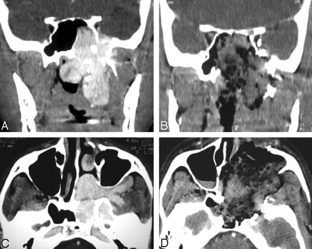
Fig 1.
True-negative result on CT scans.
A and C, Preoperative coronal (A) and axial (C) images show JNA.
B and D, Postoperative enhanced coronal (B) and axial (D) images illustrate a normal aspect of early postoperative CT when no RD is identified. There is no enhancement at the preoperative location of JNA. Hemostatic material mixed with blood is seen in the postoperative cavity.

