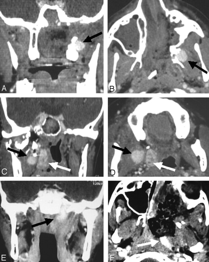
Fig 2.

True-positive result. Early postoperative enhanced helical CT reveals RD.
A and B, Coronal (A) and axial (B) images of show RD on the lateral edge of the pterygoids (arrow).
C and D, Coronal (C) and axial (D) images show RD in the pterygoid muscles (arrows).
E and F, Coronal (E) and axial (F) images show RD in the foramen lacerum (arrow).
