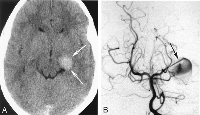
Fig 1.
A, Initial CT scan. Axial CT scan shows hyperattenuated round lesion (arrows) adjacent to the left ambient cistern with slight compression of the brain stem. B, Initial arteriogram. Left vertebral arteriogram, anteroposterior view, shows a contrast medium “jet” filling (arrow) of a large aneurysm arising from the left P2 segment.

