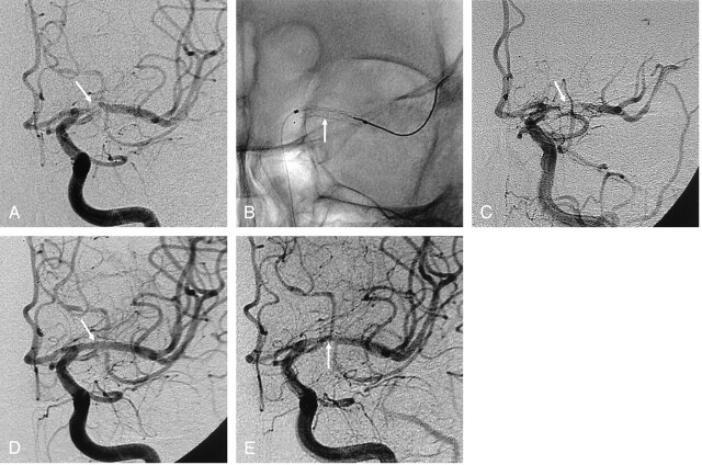
Fig 2.
A 67-year-old man with recurrent TIA symptoms.
A, Anteroposterior left ICA angiogram shows severe stenosis (about 90%, arrow) in the proximal M1 portion of the left MCA.
B, Balloon-mounted coronary stent (arrow) is successfully deployed.
C, Postprocedural ICA angiogram shows acute in-stent thrombosis (arrow).
D, Final ICA angiogram after thrombolysis with abciximab shows the MCA (arrow) with a smooth appearance, normalized luminal diameter, and preservation of the lenticulostriate arteries.
E, Angiogram at 10 months after the procedure shows asymptomatic, mild (20%) in-stent restenosis (arrow) due to intimal hyperplasia.

