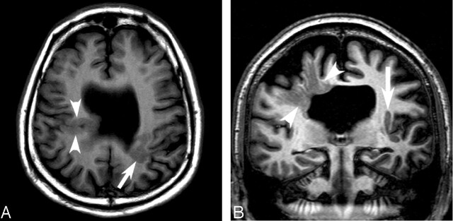
Fig 1.
Conventional MR images obtained at 1.5 T and 3 T.
A, Axial T1-weighted image obtained at 1.5 T shows bilateral closed lip schizencephaly (arrow and arrowheads) and absent septum pellucidum.
B, Coronal image obtained at 3T through the schizencephalic cleft reveals individual linear radiations of gray matter signal (arrowheads) extending from the cortex to the ventricle.

