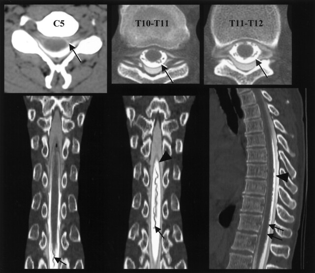
Fig 2.
Large epidural pseudomeningocele and dilated posterior thoracic spinal vein. Axial CT myelogram of the cervical and thoracic spine shows dura marginating the epidural pseudomeningocele (highlighted black arrows). Coronal and sagittal reformatted CT myelogram images of the thoracic spine demonstrate a tortuous, dilated posterior thoracic spinal vein (black arrows) and dura (arrowheads) separating intradural and epidural CSF.

