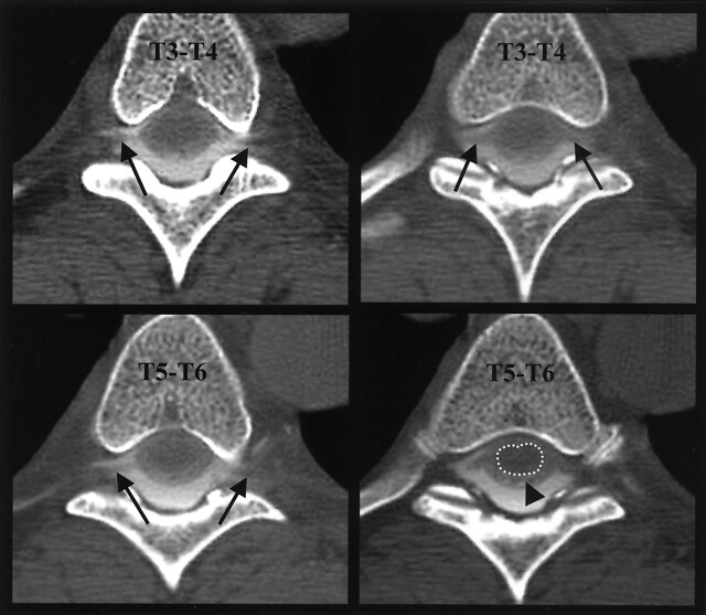
Fig 3.
Large epidural pseudomeningocele. Axial CT myelogram of the midthoracic spine demonstrates an epidural pseudomeningocele extending into the neural foramina, outlining thoracic nerve roots (e.g., black arrows). Note faint opacification of the compressed subarachnoid space (black arrowhead) surrounding the cord (dotted outline), best seen at bottom right image.

