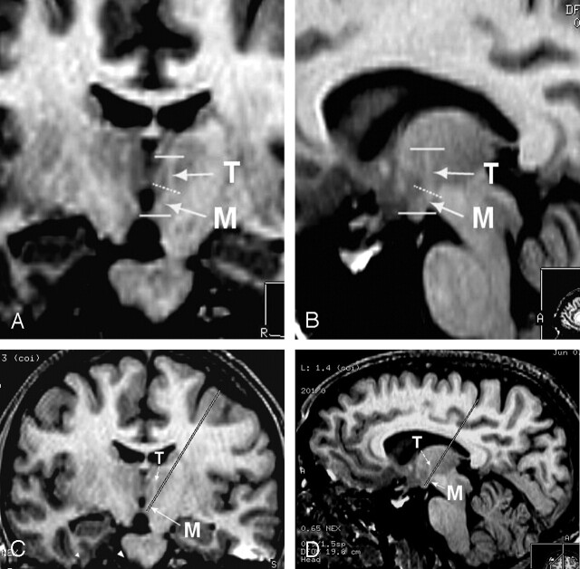
Fig 1.
Anatomic study on healthy volunteers (reformatted images from unenhanced 3D T1-weighted SPGR acquisition).
A, Coronal oblique reformat (magnified view) clearly depicts the two segments of the MTT on both sides. The presence of a proximal “mammillary” (M) and of a distal “thalamic” (T) segment separated by an angulation (dotted line) is clearly shown.
B, Sagittal oblique reformat also shows the two segments of the left MTT in an orthogonal plane relative to that in the previous illustration.
C, Electrode trajectory simulation on frontal oblique reformat (similar but demagnified view as 1A) shows “catheterization” of the mammillary segment of the MTT and safe cortical entry point located in the posterior third of the middle frontal gyrus.
D, Electrode trajectory simulation on sagittal oblique reformat (similar but demagnified view as in 1B) showing “catherization” of the proximal MTT, but critical central sulcus entry point.

