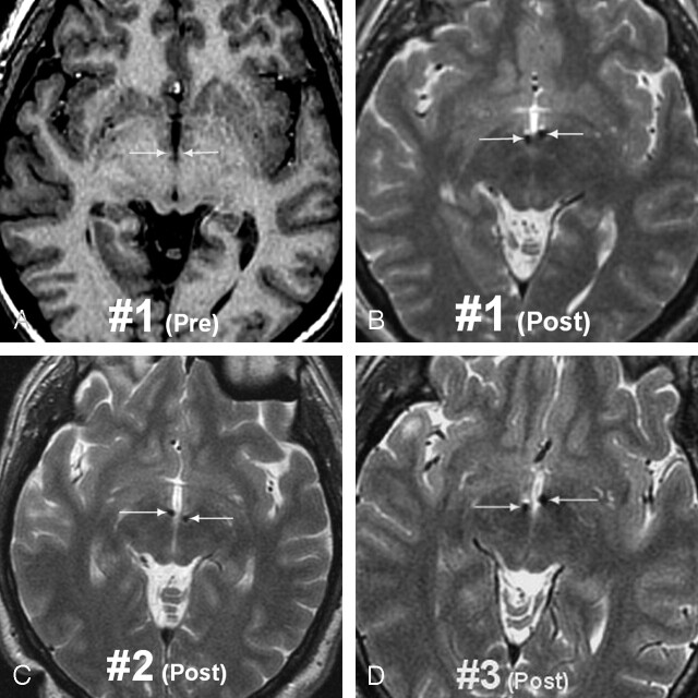
Fig 2.
MR images in the three bilaterally MB implanted patients
A, Preoperative status in patient 1 (T1-weighted 3D SPGR image) showing the mammillary segment of the MTT on both sides (arrows).
B, Postoperative image (T2-weighted 3D fast spin-echo [FSE] image) in patient 1 showing the electrodes through the mammillary segment (arrows). FSE T2 weighting was preferred to minimize susceptibility artifacts due to ferromagnetic components of the electrodes.
C and D, Postoperative status in patients 2 and 3 (T2-weighted 3D FSE images) also showing “catherization” of the mammillary segment of the MTTs in both cases.

