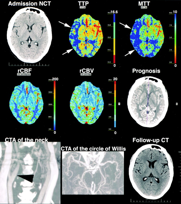
Fig 2.

77-year-old woman with acute-onset left hemiparesis. Admission nonenhanced CT 2 hours after onset is normal. TTP and MTT are prolonged in the right superficial MCA territory (arrows). rCBFs are normal, but rCBVs are higher than contralateral values. TTP and MTT changes are not explained by any vascular abnormality; right carotid bifurcation (arrowhead) and intracranial arteries are normal. Final diagnosis was TIA. Follow-up CT 14 days later was normal. TTP and MTT changes were regarded as false-positive and most likely related to luxury perfusion due to TIA.
