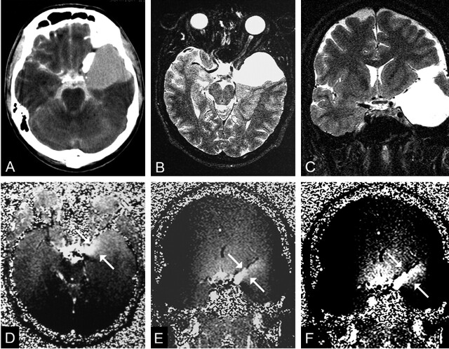
Fig 1.
A 20-year-old man with headaches and a communicating left middle cranial fossa AC (patient 10).
A, CTC shows homogeneous contrast enhancement in the cyst.
B and C, Transverse (B) and coronal (C) T2-weighted images (TR/TE/NEX, 7400/115/1) obtained before PC cine MR imaging helps in proper section orientation.
D and E, Transverse(D) and coronal (E) PC cine MR images (TR/TE/flip angle, 70/15.8/10°) show hyperintensity (arrows) arising from left chiasmatic cistern, which represents communication with the subarachnoid space.
F, Adjusting the windowing makes the flow jet (arrows) more clear.

