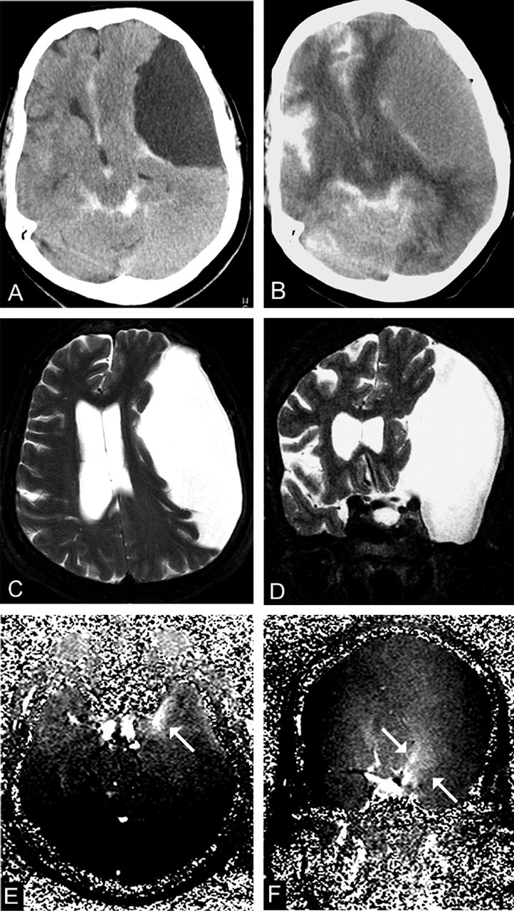
Fig 4.

A 66-year-old woman with a type III cyst in the left middle cranial fossa (patient 9).
A, CTC 2 hours after intratechal contrast injection shows no enhancement (8 HU).
B, CTC at 12 hours shows intracystic enhancement of 46 HU.
C and D, Transverse (C) and coronal (D) T2-weighted images (TR/TE/NEX, 7400/115/1) show marked midline shift.
E and F, Transverse (E) and coronal (F) PC cine MR images (TR/TE/flip angle, 70/15.8/10°) shows evidence of a flow jet (arrows), although communication with the cisternal space is unlikely in type III cysts.
