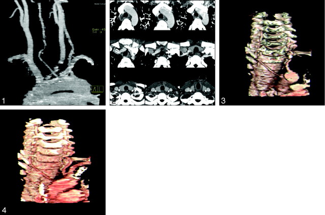
Fig 2.
Serial axial MIP images demonstrating the anomalous origin of both the vertebral arteries from the aortic arch beyond the left subclavian artery, along with the aberrant course of the right vertebral artery behind the esophagus and the trachea to reach the right paratracheal region and then to enter the right transverse foramina.

