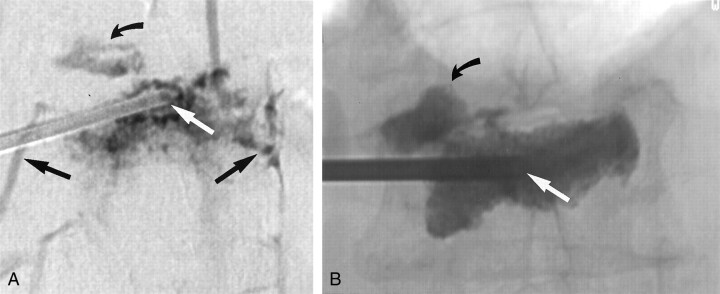
Fig 1.
Images in a 77-year-old woman with an L1 vertebral body fracture.
A, AP digital subtraction venogram shows the tip of an 11-gauge needle (straight white arrow) at the midline of the vertebral body. Multiple routes of contrast material egress are present, including routes through the superior endplate (curved black arrow) and bilateral paravertebral veins (straight black arrows).
B, AP plain radiograph obtained after vertebroplasty shows that the tip of the needle remains at the midline (white arrow). The needle position has not been altered because direct or rapid venous filling during venography was not observed. Cement fills most of the vertebral body, and it has also extravasated into the superior disk space (black arrow), in the exact same pattern as that predicted by using the venogram in A.

