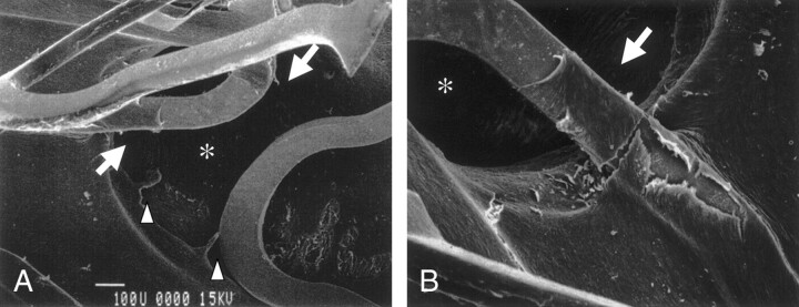
Fig 3.
Representative case (case 4). Scanning electron microscopic findings obtained 3 months after stent placement reveal an open ostium of the lumbar artery (asterisk) and also show that regenerated endothelial-like cells are covered with stent strut in contact with the aorta and extend to the struts crossing the ostium of the lumbar artery (arrows). In this case, the extension of the regenerated endothelial cells into the ostia of lumbar arteries was observed (arrow), although the degree of narrowing due to this extension was not significant.
A, Original magnification ×80.
B, Original magnification ×150.

