Fig 3.
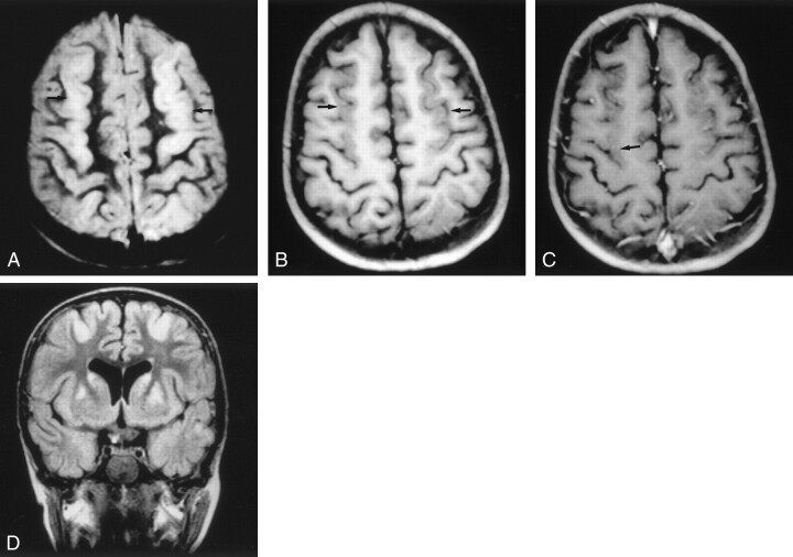
Images (MR imaging class D) of a patient who experienced hypovolemic shock and cardiorespiratory arrest due to intestinal necrosis secondary to volvulus at age 4.5 years. The outcome was good (neurologic evolution = 0).
A, Axial proton density–weighted image, obtained 18 days after the event, shows stripe of hyperintensity in the parasagittal cortex (arrows).
B, Unenhanced axial T1-weighted image (500/20), obtained 18 days after the event, shows parasagittal cortical swelling (arrows).
C, Contrast-enhanced axial T1-weighted image (500/20), obtained 18 days after the event, shows mild cortical enhancement (arrow).
D, Coronal T2-weighted FLAIR image (6000/2000/150) shows areas of hyperintense signal in the cortical parasagittal anterior region. Hyperintense signal was also seen in the posterior region (not shown) and basal ganglia (caudate and lentiform nuclei). Watershed areas score, 3/4; basal ganglia score, 3/4; total MR imaging score, 6/8.

