Fig 1.

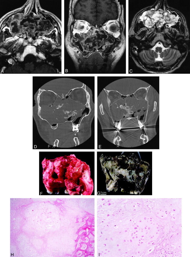
Images from the case of a 32-year-old man with chondrosarcoma involving bilateral ethmoid and maxillary sinuses.
A and B, Axial (A) and coronal (B) contrast-enhanced T1-weighted MR images (600/15/2 [TR/TE/NEX]; section thickness, 5 mm) show a large, heterogeneously enhancing mass centered at the nasal septum, involving the ethmoid sinuses and maxillary sinuses.← The mass extends into the inferior right orbit without infiltration of the rectus muscles or intraconal structures. No evidence of dural enhancement to suggest intracranial extension is present.
C, Axial T2-weighted MR image (4000/102/2; section thickness, 5 mm) shows a heterogeneous mass, as seen before, with low-signal-intensity septation.
D and E, Bone window coronal CT scans show a large lobulated mass with a calcified and chondroid matrix extending into the maxillary and the ethmoid sinuses. There is destruction of the sinus wall and the right orbital floor. D shows that the mass extends inferiorly, causing bowing of the right side of the hard palate. E demonstrates that the mass does not extend intracranially because no evidence of erosion into the cribriform plate is visible. Image in E is more posterior than image in D.
F, Bilateral maxillectomy specimen shows a tan-pink, lobulated tumor mass (6 × 4 × 4 cm) that fills the bilateral maxillary sinuses and is present at the superior (orbital) and posterior margins (arrow, not a final surgical margin). The tumor was hard to waxy, with numerous calcifications.
G, Bilateral maxillectomy specimen after decalcification shows tumor in the right maxillary sinus, extending across the nasal cavity (arrow) and into the left maxillary sinus.
H, At low power, the tumor is composed of lobules of pale-staining chondroid matrix. Infiltration of the bone is seen at the right side of the field (hematoxylin-eosin stain, original magnification ×200).
I, At higher power, the tumor resembles normal cartilage. Chondrocytes reside within lacunae in a pale-staining amorphous matrix. Compared with benign cartilage or enchondroma, however, the tumor is more cellular. Cell nuclei tend to be larger and more irregular, and binucleation within a single lacuna is more common.
