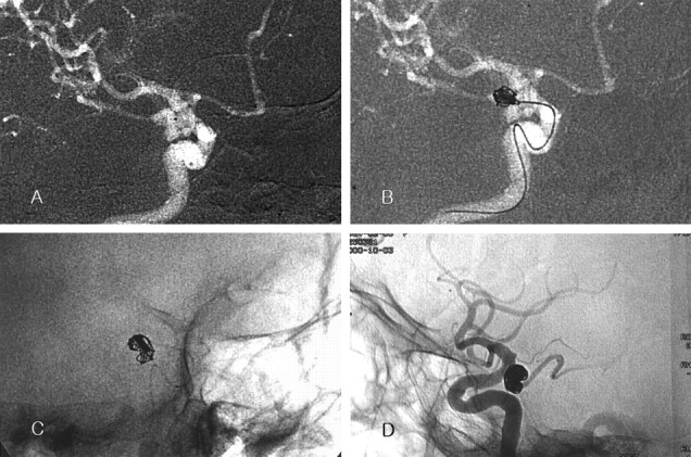
Fig 1.

Case 1. Images in a 76-year-old woman with a ruptured posterior communicating arterial aneurysm.
A, Roadmap image reveals the bilobulated aneurysm. Note the position of the catheter tip (black dot in the aneurysm).
B, The longest diameter of each lobe was 5 and 4 mm. When a 7-mm × 25-cm two-dimensional GDC-10 (GDC-10–2D) was inserted as the first coil, the coil frame did not occupy the whole inner space of the aneurysm. The coil was retrieved.
C, A 9-mm × 30-cm GDC-10–2D is inserted. The coil frame matches the contour of the aneurysm.
D, Postembolization angiogram shows compact coil packing. No contrast material fills the aneurysmal sac.
