Fig 3.
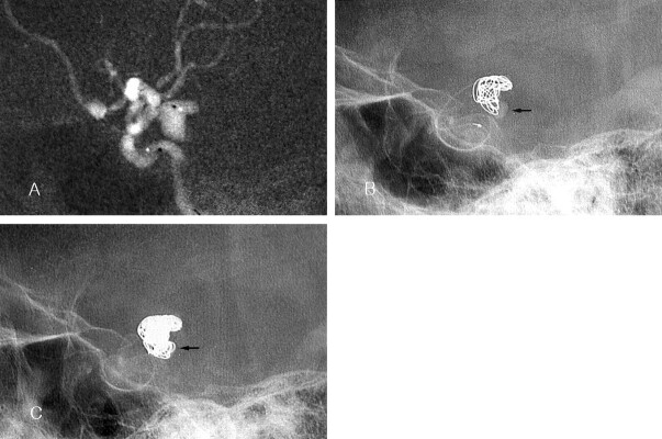
Case 3. Images in a 71-year-old woman with a ruptured left posterior communicating artery aneurysm.
A, Roadmap image shows the bilobulated aneurysm. Note the position of the catheter tip (black dot in the aneurysm).
B, The sum of the longest lengths of the two lobes was more than 10 mm. A GDC-10 coil (10 mm × 30 cm) was selected as the first coil. The frame made by the first coil does not exactly match the contour of the aneurysm. Note the contrast agent retention in the inferior lobe (arrow). Another 10 mm × 30 cm GDC-10 was placed after the first coil was detached.
C, Final fluoroscopic image shows compact coil packing. The space with contrast retention in B is occupied with coils (arrow).

