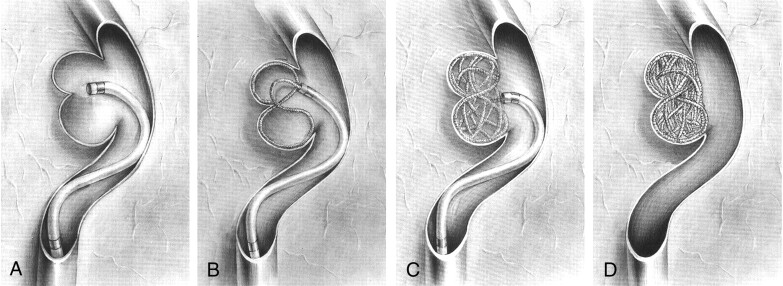
Fig 4.
Schematic diagrams of the figure-8 technique.
A, A microcatheter is positioned 1–2 mm from the isthmus of the two lobes.
B, After the first coil loop is placed into one lobe, another coil loop is made in another lobe with only further insertion. The catheter tip has a swaying movement during coil delivery. The coil loops may be shaped like a figure 8.
C, Coils are smoothly placed into one lobe and then into the other lobe with just careful further insertion. In general, the subsequent coil has a diameter 1–2 mm smaller than the previous coil.
D, Finally, the deeply bilobulated aneurysm is completely occluded.

