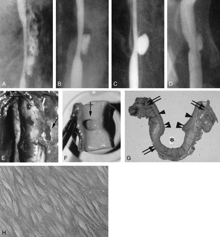
Fig 3.

Example of aneurysm formation, with persistent stenosis (group I-C 6-mm elliptical defect).
A, Angiogram shows a double shadow due to a massive adventitial hematoma that formed immediately after lesion creation.
B, Angiogram obtained 2 hours later shows a subadventitial hematoma with the characteristics of an aneurysmal pouch.
C, Angiogram obtained 1 week later shows that the hematoma has become a saccular aneurysm.
D, Chronic-stage (3-month) angiogram shows that the aneurysm is smaller.
E, Photograph shows external protrusion of the aneurysm (arrow).
F, Chronic-stage photograph obtained in shows that part of the intimal defect extends to the aneurysm orifice (arrow).
G, Chronic-stage photomicrograph shows that the inner layer of the aneurysm dome (asterisk) is covered with organized clots and fully endothelialized (elastica van Gieson stain, original magnification ×5). Arrows indicate the adventitia; single arrowheads, media; double arrowheads, intima.
H, Scanning electron micrograph shows endothelialization (original magnification ×700).
