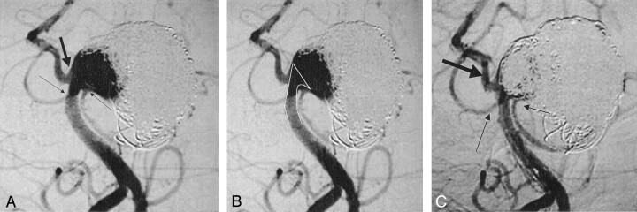
Fig 4.
A, A 54-year-old woman presenting with recurrence of a giant distal basilar artery aneurysm. The base of the aneurysm incorporates both superior cerebellar arteries (small arrows) and the right posterior cerebral artery (large arrow). The left posterior cerebral artery is occluded.
B, To maintain patency of both superior cerebellar arteries and the posterior cerebral artery, it was decided to place the stent 5 mm above the origin of the right posterior cerebral artery, directly within the aneurysm. The “white line” indicates stent deployment.
C, Following stent deployment, a total of 13 additional Orbit coils were placed above the stent and were well maintained in position by the deployed stent. The postocclusion angiogram demonstrates significant reduction in flow to the aneurysm, while maintaining sufficient blood flow to both superior cerebellar arteries (small arrows) and the posterior cerebral artery (large arrow) by the stent.

