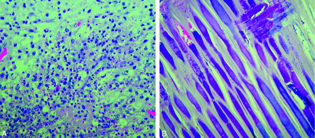
Fig 3.
Foreign body reaction.
A, Acute and chronic inflammatory changes. A mixed inflammatory infiltrate, composed of neutrophils, lymphocytes, plasma cells, and macrophages, was present in the biopsy specimen (hematoxylin-eosin, original magnification ×200).
B, Segments of foreign material (hematoxylin-eosin, original magnification ×400).

