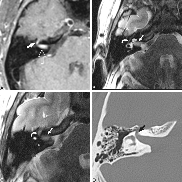
fig 3.
Dumbbell facial nerve schwannoma.
A, Axial enhanced T1-weighted MR image (800/27/2) at the level of the IAC shows an avidly enhancing mass involving the IAC (open arrow) with an enhancing “tail” involving the labyrinthine segment of the facial nerve (white arrow) as it courses toward the geniculate ganglion.
B, Axial high-resolution FSE T2 MR image (4000/130/1) at the level of the IAC shows a hypointense mass filling the IAC (white arrow) displacing the normal cerebrospinal fluid. The tympanic segment of the facial nerve is barely visible in its bony canal (curved white arrow).
C, Axial high-resolution FSE T2-weighted MR image (4000/130/1) just cephalad to B shows the enlarged labyrinthine segment mass (curved white arrow) as it courses toward the geniculate ganglion. The hypointense intracanalicular portion is again seen (white arrow).
D, Axial CT image at the level of C confirms the enlarged labyrinthine portion of the facial nerve canal (black arrow). Used by permission (37).

