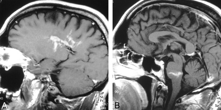
fig 1.
T1-weighted postcontrast images.
A, Left parasagittal section at the margin of the lateral ventricle. Heterogeneous, serpiginous, and linear enhancement in the lesion is well demonstrated.
B, Midsagittal image demonstrates a second contrast-enhancing lesion with a linear appearance in the lower pons.

