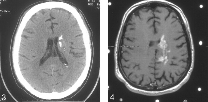
fig 3.
Noncontrast axial CT scan obtained 2.5 years after the initial imaging study. There is extensive heterogeneous calcification of the left periventricular lesion. Note the minimal mass effect, which is unchanged from previous studies.
fig 4. Axial T1-weighted postcontrast image obtained at the time of the second stereotactically guided biopsy. The lesion is similar in appearance to the earliest study

