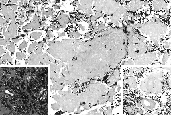
fig 5.

Masses of amyloid interspersed between fragments of reactive brain tissue (hematoxylin & eosin [H&E], magnification ×200). Right insert shows involvement of blood vessel walls with amyloid (H&E, magnification ×300), while the left shows the characteristic apple-green birefringence (Congo red, polarized ×300)
