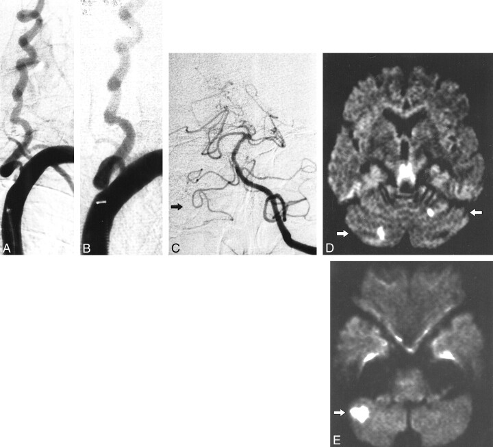
fig 3.
Patient 6.
A, Stenosis of left VA (occlusion of right VA is not shown).
B, Result after percutaneous angioplasty (3.5-mm balloon).
C, Left VA injection. Note the anterior inferior-posterior inferior cerebellar artery complex on the right side (arrow). Post-procedural diffusion-weighted images (6000/103; number of excitations, one) obtained with a diffusion sensitization level of b = 1000 s;clmm2.
D, New cerebellar lesions: 5 to 10 mm, superior cerebellar artery territory (arrows).
E, New cerebellar lesion: 20 mm, posterior inferior cerebellar artery territory (arrow).

