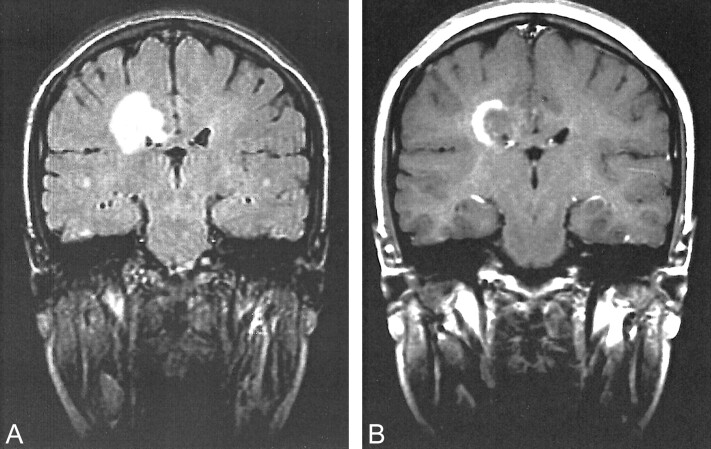
fig 2.
Images of a 20-year-old woman who was admitted because of acute left hemiparesis (case 2).
A, Coronal view fluid-attenuated inversion recovery image (10000/102/1; inversion time, 1800 ms) shows the concentric lesion with two rings located in the right periventricular white matter adjacent to the corpus callosum.
B, After injection of contrast material, enhancement at the outer ring of the concentric lesion is seen on T1-weighted MR image.

