
fig 2.
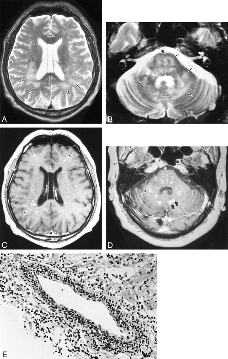
Case 2. A and B, T2-weighted images (fast spin-echo sequence with parameters of 4500/96 [TR/TE]) show diffuse hyperintense lesions in the white matter bilaterally, as well as in the pons, middle cerebellar peduncle, and cerebellar hemispheres (arrows). C and D, Contrast-enhanced T1-weighted images (spin-echo sequence with parameters 600/14 [TR/TE]) show multiple punctate and linear enhancements scattered both in the white matter bilaterally (that appear to reside along the intramedullary vessels) and in the pons and cerebellar hemispheres (arrows indicate linear enhancements). Nodular enhancement also is shown (curved arrows). E, Brain stereotactic biopsy specimen from the right occipital lobe shows inflammatory destruction of the cerebral cortex with mononuclear cell infiltration. The infiltrating cells consist of polyclonal small lymphoid cells that include nuclear atypia predominantly in the perivascular space (arrows)