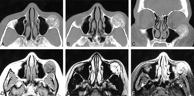
fig 1.
31-year-old woman with a 3-month history of progressive dystopia and a 1-year history of painless swelling of the left cheek.
A–C, Axial caudal (A), cranial (B), and coronal (C) CT scans, viewed at wide window settings.
A, A 2.5-cm round lesion arises from the zygoma. The inner and outer cortices, although thinned, are preserved. The trabeculae radiate in a spokewheel-type pattern, and a residual trace of the original cortex is seen (arrow).
B, The trabeculae of the lesion taper into the zygomatic arch. There is no associated soft tissue mass.
C, The rounded lesion encroaches on the inferolateral orbit.
D–F, Axial T1-weighted (500/9/2) (D), T2-weighted (3600/95/2) (E), and contrast-enhanced, fat-suppressed T1-weighted (450/9/2) (F) MR images.
D, The lesion is isointense with muscle and shows fine radiating lines of high signal intensity that are interspersed with the trabecular signal voids.
E, Encroachment on the inferolateral aspect of the orbit is seen. No extraosseous mass is evident. A line of signal representing the original cortex is visible (arrow).
F, The mass enhances; no large regional vessels are evident.

