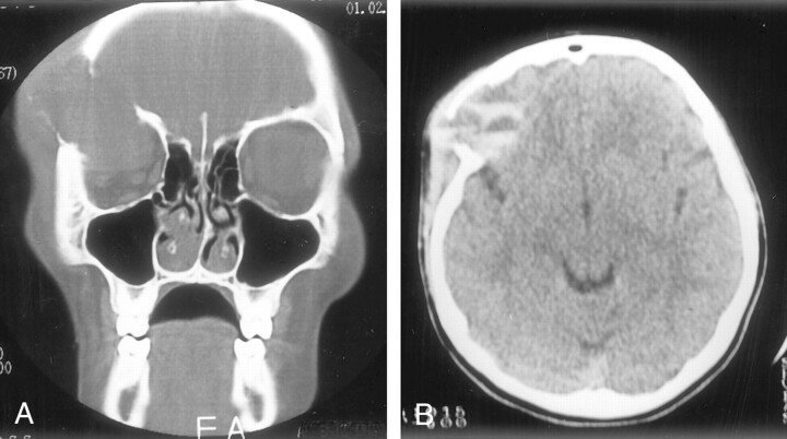
Fig 1.
Bone destruction in the superior, lateral, and posterior walls of the right orbit.
A, Coronal CT scan obtained with bone window settings shows an orbital mass causing bone destruction and displacing the orbital structures.
B, Axial non–contrast-enhanced CT scan shows multiloculated mass with fluid-fluid levels.

