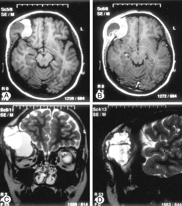
Fig 2.
Mass compressing the optic nerve and ocular muscles inferomedially.
A, Axial T1-weighted MR image (562/14/2 [TR/TE/excitations]), obtained before the administration of contrast material, shows a multiloculated mass in the right frontal region, which contains fluid-fluid levels with variable signal intensity.
B, Axial contrast-enhanced T1-weighted MR image (562/14/2) shows enhancement of the cyst walls.
C, Coronal T2-weighted MR image (4000/120/3) shows displacement of the optic nerve and ocular muscles by the mass.
D, Sagittal T2-weighted MR image (4000/120/3) shows multiple small cysts (diverticula) projecting from larger cysts.

