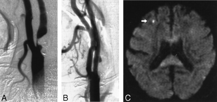
Fig 1.
Images obtained in a 70-year-old man with an asymptomatic stenosis of the carotid artery.
A, Right anterior oblique angiogram shows a 94% stenosis of the right ICA.
B, Right anterior oblique angiogram shows the result after stent implantation.
C, Postprocedural axial diffusion-weighted MR image (6000/103/1) shows a new ipsilateral lesion (<5 mm) in the cortical territory of the MCA (arrow).

