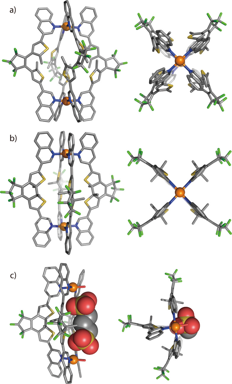
Figure 3.

X-ray structures of (a) o-C, (b) c-C, and (c) host–guest complex G1@c-B (fourth PdII positions coordinated by H2O) in side (left) and top (right) views. All hydrogen atoms, solvent molecules, counteranions, and minor disorders are omitted for clarity. In c-C and G1@c-B, all backbone methyl groups are disordered over both possible positions with approximately 50% occupancy (here only one diastereomer is shown; C gray, N blue, F green, S yellow, O red, Pd orange).
