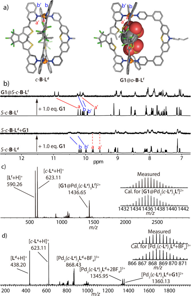
Figure 6.

(a) DFT-optimized structure of c-B-Ld (left) and X-ray structure of G1@o-B-Lf (right). (b) 1H NMR spectra (500 MHz, CD3CN, 298 K) of homochiral S-c-B-Ld and S-c-B-Lf, and their corresponding spectra after addition of 1.0 equiv of G1. (c, d) ESI-MS spectra of (c) G1@o-B-Lf, with isotope pattern of [G1@Pd2(c-La)3Lf]2+ shown in the inset, and (d) c-B-Ld after addition of 1.0 equiv of G1, with isotope pattern of [Pd2(c-La)3Ld + BF4]3+.
