
Fig 2.
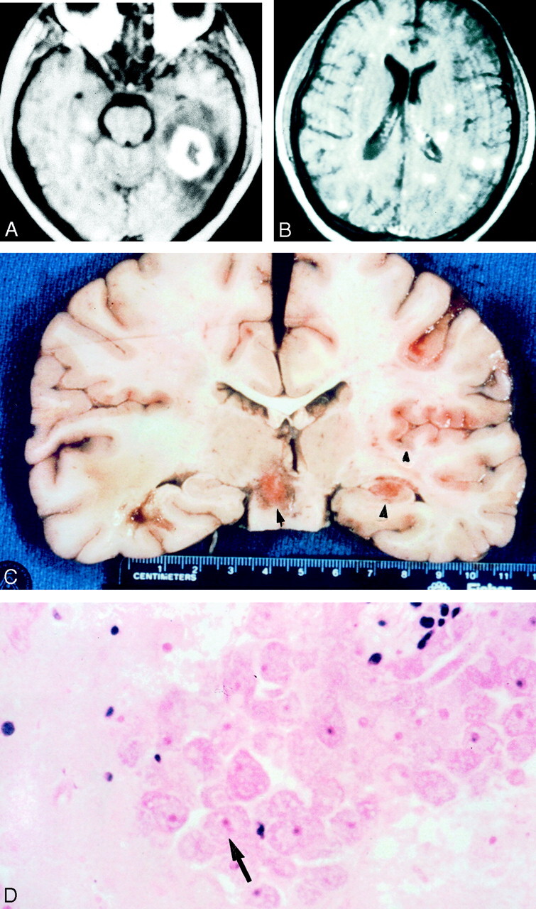
Images obtained in a 32-year-old HIV-negative man with meningoencephalitis caused by Balamuthia mandrillaris.
A, Axial T1-weighted gadolinium-enhanced MR image reveals a 3-cm enhancing left temporal lobe lesion with surrounding edema.
B, Axial T1-weighted gadolinium-enhanced MR imaged obtained on day 7 reveals innumerable enhancing areas of encephalitis that involve both gray matter and white matter.
C, Coronal gross specimen reveals multiple areas of involvement. Note the focal hemorrhages.
D, Photomicrograph reveals perivascular clustering of amebic trophozoites (arrow) (hematoxylin-eosin).