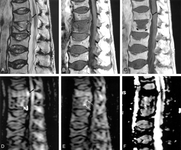
Fig 1.
Images obtained in an 83-year-old man with bacteremia and back pain.
A, T2-weighted sagittal MR image (3000/103) shows a small ventral extradural mass (upper arrow) and a fracture and abnormal signal intensity adjacent to the superior endplate of the L1 vertebral body (lower arrow).
B and C, T1-weighted sagittal MR images (815/30) obtained before (B) and after (C) IV administration of gadolinium-based contrast material show central nonenhancement in the extradural mass (long arrow in C) and in a substantial portion of the superior portion of the L1 vertebral body (short arrows in C). Both areas of nonenhancement were interpreted as abscesses.
D, Sagittal diffusion-weighted MR image (1215/80/2, b = 500) obtained at the same location as in A–C shows very high signal intensity within the extradural abscess (straight arrow) and high signal intensity within the region of the vertebral abscess (curved arrow).
E, Sagittal diffusion-weighted MR image (1215/80/2) obtained immediately to the left of D shows high signal intensity in the vertebral abscess (arrows).
F, ADC map obtained at same location as in D shows the dark appearance of both epidural (solid arrow) and vertebral abscess (open arrows) regions.

