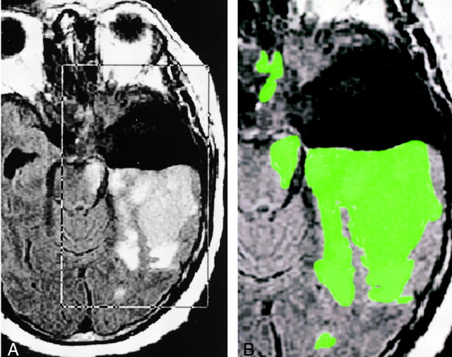
Fig 1.
Images illustrate step 2: segmentation of FLAIR images.
A, Placement of rectangular VOI around the area of presumed tumor and edema (FLAIR volume) on axial FLAIR images designated IF.
B, Delineated FLAIR volume displayed as a green overlay obtained after the deposition of seed points in the VOI.

