Fig 2.
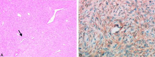
Photomicrographs in a 49-year-old woman with a bulging mass in the left oral cavity.
A, Image of the parapharyngeal mass shows that the tumor has the alternating pattern of hemangiopericytomatous area (arrow) with a hypocellular fibrous region (ie, patternless pattern); these features suggest an SFT (hematoxylin-eosin stain, original magnification ×40).
B, Image shows that the neoplastic cells have a positive reaction with CD34 immunohistochemical staining; this finding clarifies the diagnosis of SFT (original magnification ×200).

